
21 Aug Why Every Oral Mass Deserves Attention
Lumps and bumps in a pet’s mouth are often not found as readily as those on their skin by pet owners. Unless pet owners are brushing their pet’s teeth every day or happen to see oral tumors while their dog is panting, these are often found once they have grown quite large. Just by looking at an oral mass, it is impossible to tell if they are benign or malignant, so it is important to get these worked up as soon as possible.
Benign vs Malignant Oral Tumors in a Dog or Cat
Benign oral tumors do not spread to other parts of the body, while malignant tumors can spread via the bloodstream or lymphatic system to distant parts of the body such as lymph nodes, the lung or organs in the abdomen. Benign tumors often carry an excellent prognosis due to not having the potential to spread, while malignant tumors can have a variable prognosis depending on how early they were diagnosed, their size, if they’ve already spread and their position within the oral cavity. Benign doesn’t mean these tumors should be left alone. Some benign tumors may not spread but can be locally aggressive and invade adjacent tissue. Also, if benign tumors are left alone, some can grow large, causing the pet to bite into them, cause pain, difficulty eating, or the inability to fully close or open the mouth.
Meet Jax
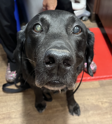
Image 1: Jax at the time of his biopsy procedure. There is a slight swelling below his left eye.
Jax is a 9-year-old Labrador Retriever that presented for an oral tumor of his upper left jaw in the back of his mouth. Jax’s owner noticed that he started having bad breath and blood-tinged drool. Jax’s primary care veterinarian found an oral tumor during his physical exam and referred Jax to Animal Dental Care and Oral Surgery.
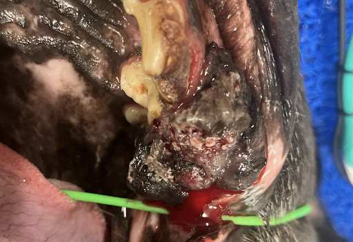
Image 2: Jax’s tumor was diagnosed as malignant melanoma.
Initial Diagnostics
Diagnostic tests are needed to determine what type of tumor is present, where it extends to in the oral cavity and surrounding tissues, and if there is any spread to any other parts of the body. An incisional biopsy is needed to determine what type of tumor we’re dealing with. An incisional biopsy means we are taking part of the tumor to send to a pathologist at an external laboratory for histopathology, but we are not trying to remove the whole tumor. Different tumors require removing different amounts of normal/healthy-looking tissue around the tumor to get the best chance of removing all of it. If you were to remove a tumor without this step, you could remove too little tissue and not get the entire tumor, or you could remove more than was needed.
Imaging is needed to determine the extent of the tumor. Imaging may include cone beam computed tomography (CBCT) or conventional computed tomography (CT). Both of these are 3-dimensional imaging modalities. Skull X-rays and dental X-rays are less useful for determining the extent of tumors because they are 2-dimensional imaging methods that have overlap of structures, which can make them difficult to accurately interpret.
CT can also be used to stage the tumor, meaning looking for spread to places such as lymph nodes and the lungs. Chest x-rays are another option for this but are less sensitive and may miss smaller changes that CT can pick up. Lymph nodes can also be aspirated or biopsied to determine if the tumor has spread to them.
Jax had an incisional biopsy of his tumor, which diagnosed a malignant melanoma, the most common malignant oral tumor in dogs. He also had a conventional CT performed of his head, neck and chest to determine the extent of his tumor and to see if there was any spread of the tumor. His mandibular lymph nodes were also aspirated to look for spread. The tumor was determined to be a good candidate for surgery, and the CT and lymph node aspirates found no signs of spread.
Definitive Surgery
If the tumor is determined to be a good candidate for surgery, a second procedure is then scheduled. This is a much larger surgery than the initial biopsy, and often includes removal of bone, such as a mandibulectomy (removal of part of or all of one of the mandibles – lower jaws) or maxillectomy (removal of part of one or both of the maxillae – upper jaws) procedure. These procedures are more invasive and painful, so dogs and cats will usually spend at least one night in the hospital for pain control. Depending on what part of the mandible or maxilla is removed, there may be cosmetic changes after the procedure, but often times these are barely noticeable and considered acceptable by most owners.
Jax had a caudal maxillectomy to remove part of his maxilla towards the back of his mouth. He stayed in the ICU overnight for pain control and was able to go home the next day with pain medications being sent home. After two weeks, Jax came back in for a recheck and was doing great at home. The biopsy results from the second procedure confirmed that the tumor was completely removed!
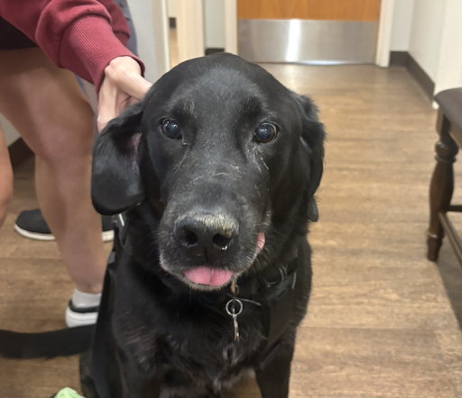
Image 3: Jax 2 weeks after his caudal maxillectomy.
After Surgery
For many oral tumors in a dog or cat, surgery is often the treatment option that leads to the best outcomes. There are additional treatment options such as radiation therapy or chemotherapy that can increase the survival time of dogs and cats with malignant tumors. These can be used in conjunction with surgery or in place of surgery if a tumor is determined to be non-surgical. A consult with an oncologist is often recommended.
If your dog or cat was diagnosed with an oral mass or if you find one at home, you can schedule a consult with one of our board-certified veterinary dentists.
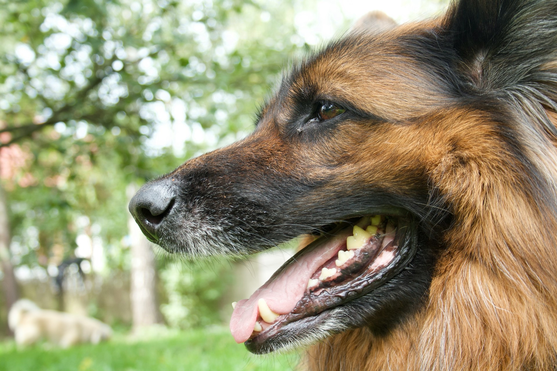
Photo by Antek on Unsplash used with permission under the Creative Commons license for commercial use 08/21/2026