01 May What is An Oronasal Fistula in Dogs and How Do We Repair it?
What is an oronasal fistula (ONF)?
By definition, a fistula is a communication between 2 different spaces. Think about a fistula like a hole in a wall that allows you to see through the wall into the next room. Oronasal fistulas are abnormal communications, or holes, between the oral cavity and nasal passageway.
When looking at the normal anatomy of the upper jaw (maxilla), the nasal passageways are separated from the oral cavity by soft tissue and bone (like a wall). When these tissues are damaged a communication can be created, like a hole in a wall, between the oral cavity and nasal passageways.
How do dogs and cats get oronasal fistulas?
The most common cause of oronasal fistulas is periodontal disease. Periodontal disease is inflammation of the supporting tissues around the teeth. These tissues include the gingiva, periodontal ligament, cementum and bone, all anchoring the teeth in place. As periodontal disease progresses, these tissues are eroded away and lost. With enough erosion, the periodontal tissues become weakened or completely eroded away to form a fistula between the oral cavity and nasal passageways. The most common site for an oronasal fistula is along the upper canine (fang) teeth. However, they can form around other teeth as well.
Other common causes of oronasal fistulas are trauma (getting hit by car, bit by another dog, suffering a penetrating wound, etc.) to the maxillofacial area (upper jaw), aggressive oral cancers, and at times during a tooth extraction. The bone separating the tooth root and nasal passageway is thin around some roots. If over-exuberant extraction techniques are used or if the bone is already weakened due to periodontal disease, the bone can break creating an oronasal fistula.
How do I know if my pet has an oronasal fistula?
If the oronasal fistula is large enough, it may be seen during an awake oral exam. Otherwise, oronasal fistulas can be challenging to diagnose unless the patient has a thorough exam under anesthesia. The majority of oronasal fistulas are suspected based on clinical symptoms of rhinitis, such as nasal discharge and sneezing or in patients with advanced dental disease.
How do we treat oronasal fistulas?
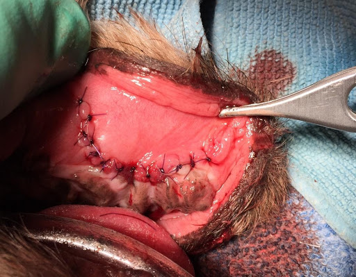
Oronasal fistulas must be surgically corrected. Once located, the fistula is sutured closed in order to reestablish the separation between the oral cavity and nasal passageway, much like patching a hole in the wall. The surgical repair includes creating a flap from oral tissues and suturing the flap over the fistula to repair it (see Figure 2 and Figure 3 below).
If a fistula is associated with a tooth, the tooth is extracted, and the site is sutured closed. For most oronasal fistulas the recovery time is the same as a tooth extraction. The patient is fed soft food and not allowed to chew on anything hard for 2 weeks or longer while the surgery site heals.
Figure 1. Intraoral picture of an oronasal fistula in a dog. Note the hole next to the canine tooth. The hole is an oronasal fistula and is a direct communication between the oral cavity and the nasal passageway. These can be difficult to diagnose in an awake patient. This picture was taken while the patient was anesthetized.
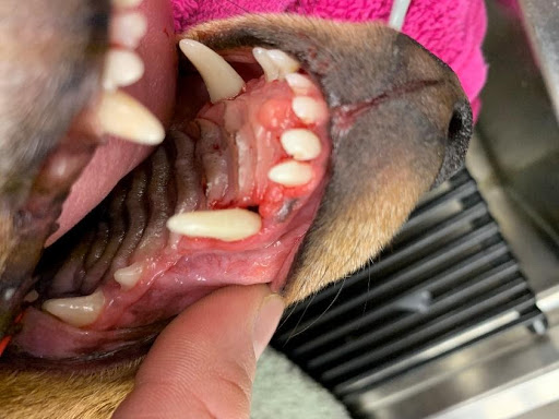
Figure 2. Intraoral picture of an oronasal fistula in a dog. Note the hole in the location where the right upper canine tooth once was. This is a larger opening to a fistula and may be diagnosed when the patient is awake.
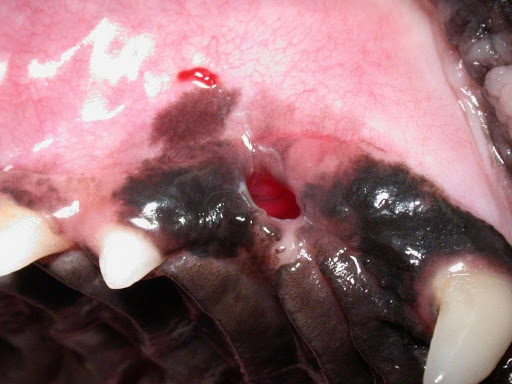
Figure 3. Intraoral picture of an oronasal fistula after surgical repair. The fistula was located where the upper right canine tooth once was. A flap of oral mucosa was sutured in place over the oronasal fistula.