
17 Jun How Can Board-Certified Veterinary Dentists Save Dog Teeth?
The oral cavity is the first part of the digestive tract and is an essential part of the digestive function of dogs and cats. Additionally, teeth-bearing animals use their teeth not only for eating but also for grooming, playing, hunting, and protection. A common goal of veterinary dentists is to preserve teeth whenever possible. Similar to human dentists, veterinary dentists have a full arsenal of techniques and materials to preserve teeth always with the goal of alleviating pain. Common procedures veterinary dentists utilize to save teeth include root canal therapy, open root planing, guided tissue regeneration, and restoration procedures.
Root Canal Therapy
Root canal therapy is most often completed in fractured or discolored teeth that are no longer vital (alive). Root canal therapy is an endodontic procedure. Endo means inside, and root canal therapy treats the innermost portion of the tooth, which is the pulp cavity. Root canal therapy preserves the tooth structure’s function, although the tooth is no longer alive. The pulp tissue is removed, and the tooth is irrigated to remove infection, followed by the placement of filling and restoration materials.
The most commonly treated teeth with root canal therapy are the canine (fang) teeth and carnassial teeth. These are strategically vital teeth that perform the most important functions in an animal’s mouth. The great benefit of root canal therapy is that it saves a pet from having to go through a surgical extraction. The structure and function of the tooth are preserved and the recovery time is minimal. Root canal treatments performed by a board-certified veterinary dentist have an excellent long term prognosis.
Root Planing & Guided Tissue Regeneration
Closed root planing procedures treat mild attachment loss of the tissues surrounding the tooth. Attachment loss begins when plaque and calculus accumulate at the gumline. Calculus accumulation at the gumline incites gingivitis and progresses to periodontitis if left untreated. Periodontitis is periodontal disease that goes beyond the gingiva. Tissue attachment decreases, leading to an increased pocket depth between the gingival tissue and bone (think of an unzipped coat). Bacteria accumulate within the pocket, causing a progression of periodontal disease. If attachment loss is detected at an early stage, the exposed root surface can be cleaned, followed by the application of a specific antibiotic gel, which promotes reattachment of the periodontal tissue and halts the progression of periodontal destruction.
Moderate attachment loss of the tissues around a tooth requires more aggressive tactics. Open root planing with guided tissue regeneration is a procedure that serves this purpose. In guided tissue regeneration, a gingival flap is created to expose the root surface for cleaning and debridement due to the depth of the attachment loss. Once the infected tissue is removed and the root surface meticulously cleaned, different types of bone graft material are placed in the deep pocket and the gingival flap is surgically closed. This procedure allows for osseous (bone) regeneration around the root structure and regrowth of the periodontal ligament. This procedure also carries an excellent long term prognosis when performed by a board-certified veterinary dentist.
Restoration Procedures
Restoration procedures involve repairing structural damage to the tooth that does not include pulp exposure. Common tooth pathologies requiring restoration procedures include carious lesions and enamel dentin fractures. In caries, also known as cavities, the carious tissue is removed, followed by placement of restoration material. This is important since without treatment the lesion will progress, leading to the eventual loss of the tooth. Enamel dentin fractures are commonly referred to as uncomplicated crown fractures that do not result in direct pulp exposure. An odontoplasty (smoothing) and bonded sealant procedure is used to treat these cases. The fractured site is smoothed, followed by the application of a dentin bonding agent and unfilled resin to protect the tooth. Crown fractures and caries are painful. When left untreated they will often lead to internal infection of the tooth causing even more pain.
Board-Certified Veterinary Dentist in Colorado Springs, Castle Pines and Loveland
Preventative care in the form of daily brushing and annual dental cleanings are essential for maintaining your dog’s oral health. All of the procedures mentioned above are performed with the goal of relieving pain and infection in your dog’s oral cavity. Colorado is such a beautiful place to live and be outside with your pets. Don’t let dental pain keep them “under the weather” and inside! At Animal Dental Care and Oral Surgery in Colorado Springs (with offices in Castle Pines and Loveland), our mission is to provide a pain-free mouth for our patients and to save their teeth whenever we can. Give us a call today to schedule preventative or restorative treatments.
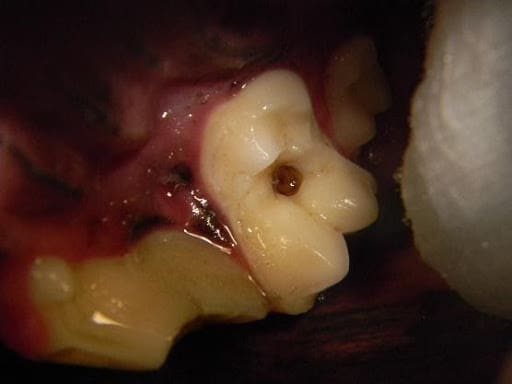
Figure 1. A maxillary molar in a dog with an active caries (cavity) lesion prior to treatment
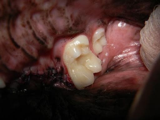
Figure 2. The same tooth after treatment with a composite restoration. The tooth to the left had a severe crown fracture that required surgical extraction.
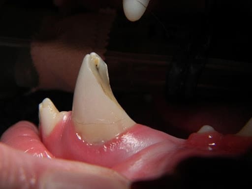
Figure 3. A canine (fang) tooth in a dog with a painful and infected crown fracture with pulp exposure
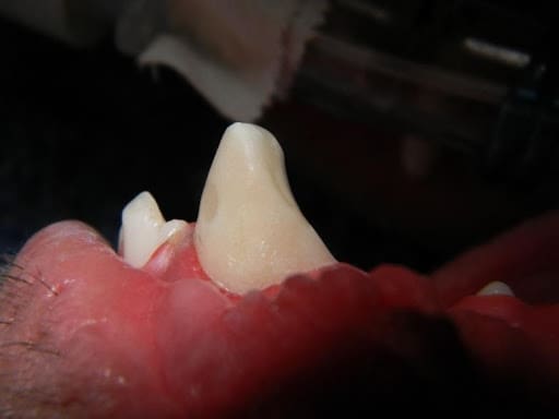
Figure 4. The same tooth after root canal therapy and crown restoration was performed.
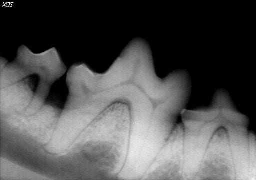
Figure 5. A large mandibular molar in a dog with severe bone loss from periodontal disease. Unfortunately, the small molar to the left could not be saved and was surgically extracted.
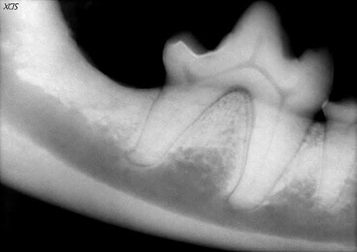
Figure 6. The same tooth 6 months after an open root planing and guided tissue regeneration procedure. The lost bone and periodontal ligament have been regenerated.