29 Jun Common Dental Problems Leading to Tooth Extraction in Dogs & Cats
There are numerous reasons why a dog or cat tooth would need to be extracted. Some of the dental problems leading to tooth extraction can be prevented, or at least minimized, and some cannot. Our goal is to always save teeth when possible, however, there are circumstances when a disease process is too advanced for a tooth to be saved. Some of the common indications for tooth extraction include advanced periodontal disease, tooth fracture, endodontic disease, tooth resorption, and caries/cavities. Ultimately, extraction is always better than leaving a painful, diseased tooth untreated in the mouth.
1. Periodontal Disease
Periodontal disease is one of the most common disease processes identified in dogs and cats. It results from the immune system’s inflammatory reaction to plaque bacteria that leads to the destruction of periodontal tissues. Periodontal disease starts in the gingiva. Inflammation of the gingiva (gingivitis) can progress to inflammation of deeper tissues that support the tooth. As periodontal disease progresses the attachment loss around the tooth worsens. Loss of periodontal attachment can rapidly progress to the point where the tooth cannot be saved and extraction is indicated.
2. Complicated Crown Fractures (Pulp Exposure)
Complicated crown fractures are tooth fractures that expose the pulp (blood vessels and nerves) of the tooth. Complicated crown fractures are painful and are always considered to be infected and dead or dying. Tooth fractures occur often when our pets chew on hard objects (i.e. rocks, bones, antlers, or other hard toys) or have an unforeseen trauma to the mouth. These fractured teeth should never be just monitored. They should always be treated with either root canal therapy or extraction as soon as possible. Root canal therapy spares the dog or cat from having to go through a surgical extraction.
3. Uncomplicated Crown Fractures and Non-vital Teeth
Uncomplicated crown fractures and blunt trauma to the tooth are common causes of endodontic disease. Uncomplicated crown fractures (“chip” fractures) are fractures involving the enamel and underlying dentin, but do not extend into the pulp chamber. Uncomplicated crown fractures are often identified during awake or anesthetized oral exams. Although the pulp is not directly exposed, infection of the pulp is possible through the exposed porous dentin. Bacteria can migrate through dentin tubules and possibly infect the pulp.
Blunt force trauma often leads to irreversible inflammation of the pulp, discoloration and death of the tooth. In both scenarios, infection and the formation of tooth root abscesses may occur and can be a significant source of pain and discomfort affecting quality of life. The treatment of these teeth is either root canal therapy or extraction. Many uncomplicated crown fractures do not lead to death of the tooth and can be treated by smoothing the fracture site and sealing it with a bonding agent.
4. Tooth Resorption
Tooth resorption can be diagnosed in both dogs and cats. This disease syndrome results in the destruction of the tooth structure and possible nerve exposure and pain. Tooth resorption is commonly identified in cats, affecting approximately one-third of the feline population. Pets affected by tooth resorption often have behavior changes or changes in their eating habits that often go unnoticed. The recommended treatment for tooth resorption with nerve exposure is always extraction.
5. Caries (Cavities)
Caries (cavities) are most often identified on the occlusal surface of molar teeth in dogs and destroy enamel and dentin with possible pulp exposure. Cavities are caused by the bacterial breakdown of highly refined carbohydrates resulting in the production of lactic and acetic acid that erode enamel and dentin. With early diagnosis, cavities can be treated with a dental filling. If left untreated, the cavity will eventually erode through the enamel and dentin of the tooth exposing the pulp chamber. Root canal therapy may be an option if there has not been extensive destruction of the tooth, but extraction is often the only option.
Surgical Tooth Extractions in Dogs and Cats
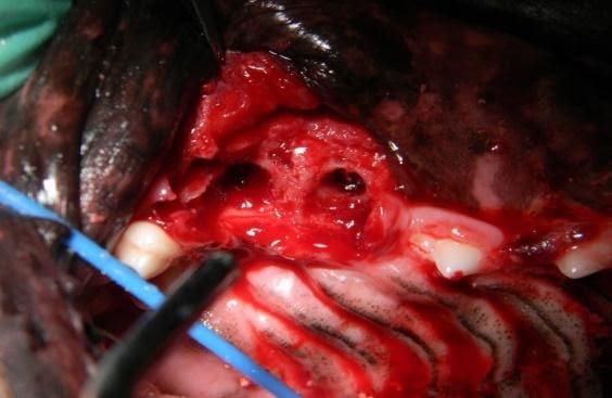
Surgical extractions begin with elevating the oral tissue away from the tooth and bone. Approximately, two-thirds of the tooth is below the gingiva and surrounded by bone. Many teeth also have multiple roots that need to be surgically separated with high-speed equipment. Some of the bone surrounding the roots often requires removal allowing the teeth to be extracted with dental elevator instruments. The underlying bone is then smoothed and the soft tissues are sutured in place over the extraction site. Possible complications of surgical extractions may include bleeding, delayed healing due to infection, and dehiscence (breakdown) of the surgery site. Surgical extractions are always painful procedures that require post-operative pain relief for 7 or more days.
Board-Certified Veterinary Dentist in Colorado Springs
Dental disease is common in dogs and cats and often progresses to the point where surgical extraction is the only option. The goal is always early diagnosis and treatment of painful dental disease hopefully before extraction is required. To hopefully avoid surgical extractions for your pet, consult regularly with your local veterinarian or a board-certified veterinary dentist to learn the best forms of dental disease prevention. If your dog or cat is suffering from dental disease or is in need of a tooth extraction in Colorado Springs, give us a call at Animal Dental Care and Oral Surgery. Colorado is such a wonderful state to live in. Let’s do all we can to keep our pets happy and healthy in our beautiful environment.
Figure 1. Periodontal disease. (A) Note the gingival recession and bone loss around the root of the upper 4th premolar. As the disease process progresses, the periodontal structures that anchor the teeth in place continue to be destroyed. (B) A more advanced, or end-stage periodontal case example. Note the loss of bone between the roots exposing this space. Surgical extraction is the only viable treatment for these teeth.
(A) 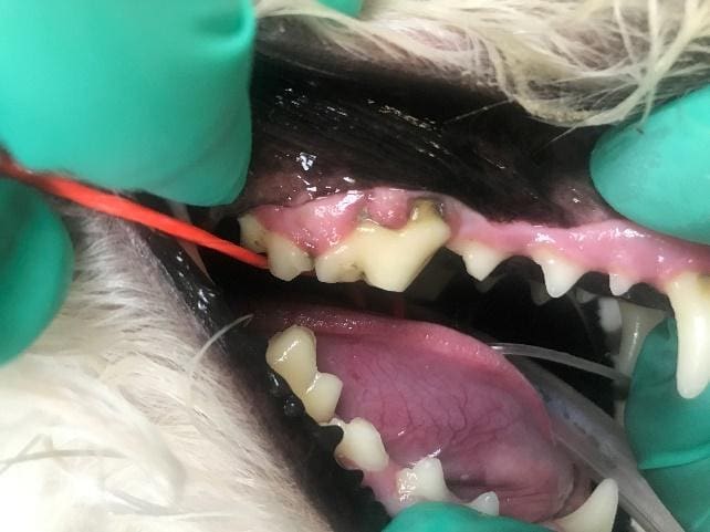 (B)
(B) 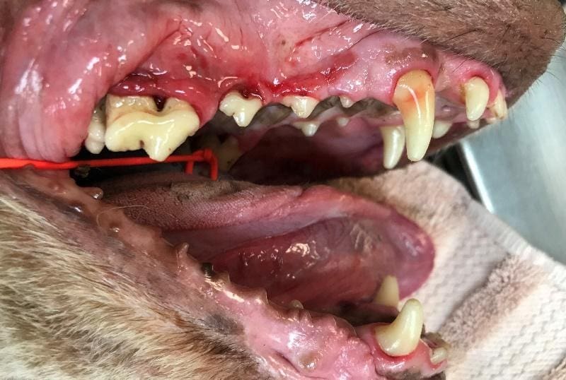
Figure 2. Two examples of complicated crown fractures that exposed the pulp of the tooth. (A) The red dot on the crown of the tooth indicates pulp exposure. (B) The start of oral surgery to extract a fractured tooth. In this step, the gingiva and oral mucosa are being elevated off the underlying bone. The bone is then removed to expose the roots.
(A) 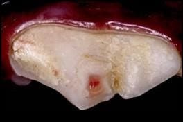 (B)
(B) 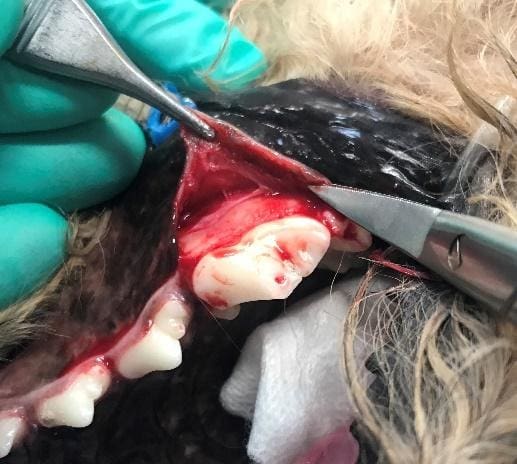
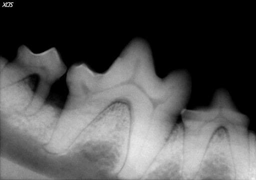
Figure 3. (A) A tooth of an uncomplicated crown fracture and (B) the radiograph of the same tooth with abscesses around the tooth roots. (C) A discolored tooth.
(A) 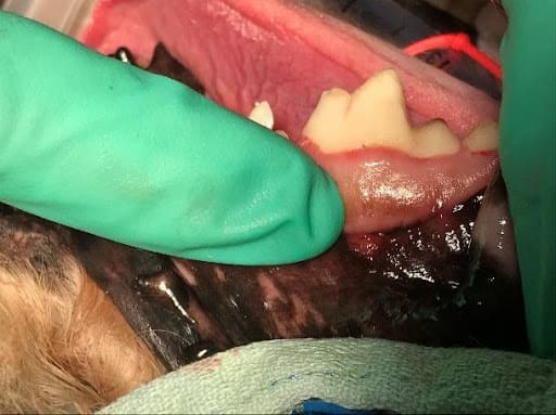 (B)
(B) 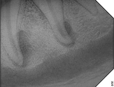 (C)
(C) 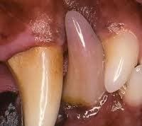
Figure 4. A feline premolar tooth with inflammatory tooth resorption.
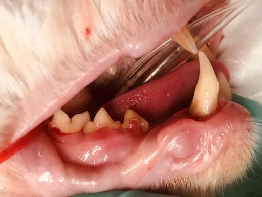
Figure 5. (A) A molar a cavity that can be treated with a restorative filling and (B) a tooth with an end-stage cavity that requires extraction.
(A) 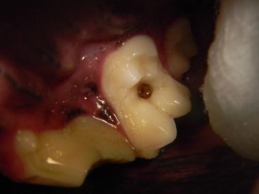 (B)
(B) 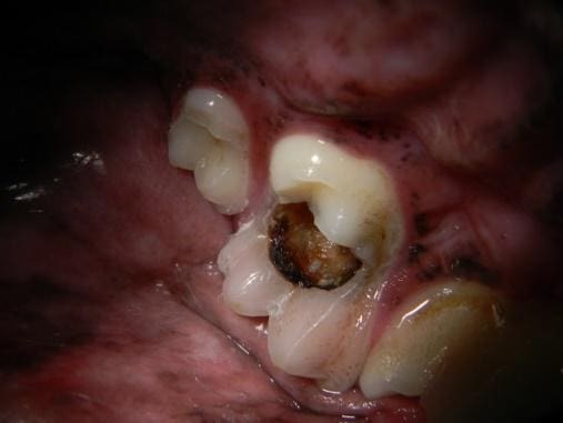
Figure 6. An extraction site after the tooth structure has been removed. Note the flap of oral tissue which has been elevated off the underlying bone to allow for bone removal.