
02 Jun Diagnosis and Treatment of Dentigerous Cysts in Dogs
What is a dentigerous cyst?
A cyst is an abnormal fluid-filled sac or space. Cysts can expand and be locally destructive. Dentigerous cysts form in the oral cavity and are commonly diagnosed in dogs, and less commonly in cats. These are cysts of dental origin that form from tissues that are a part of the tooth structure.
Dentigerous cysts form around unerupted, impacted teeth. During the eruption of a tooth into the oral cavity, the layers of the developing tooth that produce enamel, the external covering of the crown, is lost. This is normal. When teeth are impacted and remain under the gum tissue, these same layers remain present. The layers that would normally be lost during eruption of the tooth, produce fluid, resulting in cyst formation around the impacted tooth.
Where do dentigerous cysts usually form in the mouth?
Although there is the potential for these cysts to form around any tooth in a dog or cat, they occur more often in particular teeth and breeds. The most commonly impacted teeth that form cysts are the upper and lower canine teeth and lower first premolar teeth. These cysts can occur in cats, however, they occur much more frequently in dogs and particularly in brachycephalic breeds, like Boxers, Pugs, Boston Terriers and Shih Tzu’s. As dentigerous cysts expand, they may involve surrounding teeth and further weaken the surrounding bone. This can lead to loss of additional teeth and potentially jaw fracture due to weakening of the surrounding bone structure.
How are dentigerous cysts identified?
Expanding dentigerous cysts are not often identified when evaluating the oral cavity of an awake animal. The most common clue, leading to suspicion of a dentigerous cyst, is a missing tooth that failed to erupt. It is important to remember that missing teeth are not always truly missing. Often teeth fracture, leaving behind a portion of the tooth below the gum tissue. Impacted teeth that fail to erupt often lead to dentigerous cysts. For this reason, any missing tooth needs to be further evaluated.
Diagnostic imaging, with either dental radiographs or cone beam CT scan, is needed to evaluate the dental tissues below the gum line. Identification of an impacted tooth with a fluid-filled space around the crown of the tooth is most often indicative of a dentigerous cyst.
What is the treatment for a dentigerous cyst?
Dentigerous cysts are treated with surgery to remove the impacted tooth, drain the fluid and debride (scrape) the interior lining of the cyst. Thorough debridement of the cystic lining is necessary or the cyst will recur. Once the cyst has been thoroughly curetted, the bone is smoothed and gum tissue is closed over the site.
The bone surrounding the cystic area will gradually remodel to the normal anatomy. The cystic lining should be submitted for analysis by a veterinary pathologist. Dentigerous cysts are the most common result of an impacted tooth, however, oral tumors may produce cysts and have a similar appearance to dentigerous cysts.
Figure 1. Seven-year-old, female spayed Pitbull with a missing lower left first premolar tooth. A tooth may be missing or appear missing for several reasons. Reasons for a missing tooth include a failure of development, it was previously extracted, it was lost to periodontal disease, it fractured below the gum line, or the tooth is impacted and hasn’t erupted.
The patient pictured here had a missing lower left first premolar tooth. Swelling or other abnormalities were not noted on the oral exam. Cone beam CT was used to further investigate the missing tooth.

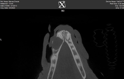
Figure 2. Below are two images from a cone beam CT scan of the above patient. These images reveal an expansile lesion in the bone of the lower-left mandible.
A) The first picture is an image looking down the length of the mandibles. There is a clear asymmetry when comparing each mandible as seen in the image. In the left mandible, there is a round lesion with a distinct border of thin bone, indicative of a dentigerous cyst.
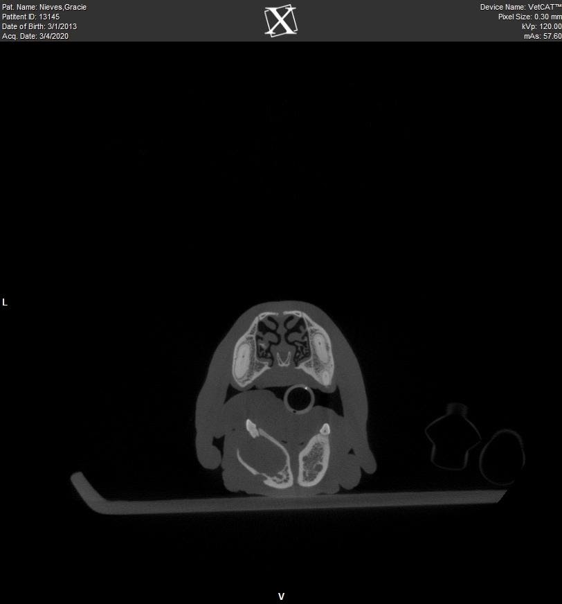
B) The second image is another view of the same lesion. This image is looking down on the mandible in cross-section. Again, the large, expansile lesion with a distinct border of thin bone.